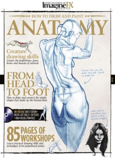
‘How To Draw And Paint Anatomy’ PDF Quick download link is given at the bottom of this article. You can see the PDF demo, size of the PDF, page numbers, and direct download Free PDF of ‘How To Draw And Paint Anatomy’ using the download button.
How To Draw And Paint Anatomy PDF Free Download

How To Draw and Paint Anatomy
Start by studying anatomy – make lots of sketches and drawings of body parts like hands, feet, arms, legs, torso, etc. This will help you understand shape, proportion, and structure. You can work from reference photos, life sketches, or anatomical sketches.
Learn Anatomy – Study bone structure, muscle structure, and how muscles attach to bones. Understand how the parts fit together and function. This will allow you to draw and paint shapes with greater precision.
Use mannequins and basic shapes – Light sketches of mannequins, cubes, spheres, etc. can help you sketch out the main shapes before adding details. This helps in keeping the ratio correct.
Focus on major muscle groups – Focus on major muscle groups like pectorals, trapezius, deltoids, abdominals, quadriceps, etc. These define the form. Minor information may come later.
Pay attention to landmark features – Pay attention to key anatomical landmarks such as the rib cage, hip bones, and collarbones. These show the proportions and location of muscles/features.
Start by studying, then focus on drawing – sketch out the body, focusing only on the arms, hands, or legs before creating a full portrait. Then incorporate what you’ve learned.
Use different contexts – Take context from different poses and multiple angles. Observing the anatomy from multiple perspectives will help in drawing/painting.
Practice smooth shading – smooth gradations in tone help to delineate rounded muscle forms. Pay attention to shadows and highlights.
fibers of attachment of the trapezius. In addition, the latissimus dorsi arises from the last three ribs as well as from the hinder end of the iliac crest for a variable distance, as has been already described.
The fibers pass upwards and outwards with varying degrees of obliquity towards the hinder fold of the armpit, the contour line of which they form:
here the muscle is twisted on itself to reach the front of the upper part of the shaft of the humerus into which it is inserted (Pls., pp. 34 38, 44 59 52, 54 94 98, 122, 126, 138, 152, 158, 162).
A triangular interval between this nucleus in the scapular regions is to be noted, the apex of which corresponds to the root of the spine of the blade bone.
Of its sides, the inner corresponds to the outer margin of the trapezius, the outer to the posterior border of the deltoid, while its base, somewhat curved, coincides with the upper border of the latissimus dorsi.
It is within the area so defined that the blade-bone, and some of the muscles connected with it, come into direct relation to the surface.
With the limbs by the side, we recognize, in this triangular interval, the internal border of the blade bone.
To the inner side of this border, we note the presence of that portion of the rhomboideus muscle which is uncovered by the trapezius.
Arising from the posterior surface of the blade bone below the spine, we see as much of the infra-spares and tenes minor as uncovered by the deltoid, while, corresponding to the lower angle and external border of the blade bone, how much of the feres major as is not overlapped by.
| Writer | – |
| Language | English |
| Pages | 117 |
| Pdf Size | 36.58 MB |
| Category | Art Book PDF |
Anatomy Drawing PDF Free Download
